Focus on Cancer RSS Feed 2014-08-06 10:00:00

Ride to Conquer Cancer Philadelphia

When we think of cancer, we don’t always think of cancer in animals. But, for Sarah Rauers, administrative assistant, for the animal biology department at the University of Pennsylvania School of Veterinary Medicine, cancer in animals – and people – is always in her thoughts.

“At this point, I don’t think there is anyone who doesn’t know someone with, or who has had cancer,” says Sarah. “I’ve had family members, as well as pets, I’ve watched suffer through the disease.”

And that, says Sarah, is one of the things that has motivated her to join the Philadelphia Ride to Conquer Cancer this October.

“I went to an information session with my co-workers, and we decided to start a team together,” says Sarah. “There was so much energy and hope we saw within the ride and the research happening at Penn, we wanted to support the cause.”

Cancer Research at Penn Vet

Within the faculty at Penn Vet, there is a lot of research that focuses on cancer. “Penn Vet studies cancer biology in mice, dogs and larger animals – all part of a mission to pursue translational medicine,” says Sarah. “Breakthroughs in cancer research today made in research could someday can one day help conquer cancer in all species.”

The other motivation behind Sarah’s decision was a very personal one.

“A couple of years ago I was hit by a car while commuting on my bike to work,” she says. “I suffered a great deal of physical and emotional trauma from that experience. Getting back on my bike and participating in The Ride is a big milestone for me.”

As for her fundraising efforts, Sarah has been pleasantly surprised at the support she’s gotten from family and friends.

“Fundraising has been so much easier than I thought it would be. Shocked by the generosity of my friends and family, and ‘Facebook friends,’” she says. “If you are considering joining, just do it. For about three days after my team signed up we were concerned about what we got ourselves into but it’s going to be easier and more rewarding than any of us think!”

Check out more from Team Penn Vet here.

The Ride to Conquer Cancer®

Ride to Conquer Cancer Philadelphia

Join Penn Medicine’s Abramson Cancer Center (ACC) on October 11 to 12, 2014 in the Ride to Conquer Cancer (RTCC)—an unforgettable and epic bike ride through Pennsylvania’s picturesque landscape -towards one life-changing destination: to cure cancer. The ride isn’t just for cyclists, it’s for anyone who wants to see a cure for cancer.

At two-days and 150+ miles the RTCC will be a physical challenge—and an emotional and inspirational weekend—that will give you a chance to ride side by side with physicians, patients and families–raising serious funds and awareness in the ACC’s fight to cure cancer.

The funds raised through the ride will be put to use immediately, powering the ACC’s vision to eradicate cancer as a cause of human disease and suffering through precision medicine, novel research, next-generation therapies, and compassionate care.

Join Today

This event will be remarkable, bringing together communities of cancer survivors, cyclists, and their supporters with a common goal to conquer this disease. Join the ride in October by registering today at www.ridetovictory.org or by calling (844) 777-7433.

Before the ride, you will have access to:

  • Expert coaching
  • Training rides in your area
  • Personal web page for fundraising
  • Helpful manual
  • 2014 ride commemorative item

During the ride, participants will have access to:

  • Event-day ride jersey
  • Support along the route
  • Catered meals
  • Entertainment at camp
  • Massage and medical care

Biomimetic Tissue-Engineered Systems in Cancer Research

Advanced technologies and biomaterials developed for tissue engineering and regenerative medicine present tractable biomimetic systems with potential applications for cancer research. Recently, the National Cancer Institute convened a Strategic Workshop to explore the use of tissue biomanufacturing for development of dynamic, physiologically relevant in vitro and ex vivo biomimetic systems to study cancer biology and drug efficacy. The workshop provided a forum to identify current progress, research gaps, and necessary steps to advance the field. Opportunities discussed included development of tumor biomimetic systems with an emphasis on reproducibility and validation of new biomimetic tumor models, as described in this report. Cancer Res; 74(19); 1–5. ©2014 AACR.

Frequently Asked Questions About Digital Breast Tomosynthesis (3D Mammograms)

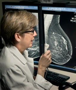
Penn researchers participated in the recently published study entitled: “Breast Cancer Screening Using Tomosynthesis in Combination with Digital Mammography,” which reviewed almost half a million mammography exams. The study, published in the June 25, 2014 issue of the Journal of the American Medical Association (JAMA), found that tomosynthesis imaging, or 3D mammography, finds significantly more invasive or lethal cancers than a traditional, 2D mammogram.

3D mammography also reduces the number of women called back for unnecessary screenings; thereby reducing anxiety and health care costs.

Specific improvements with 3D mammography screening in the research found:

  • A 41% increase in the detection of invasive breast cancers
  • A 29% increase in the detection of all breast cancers
  • Digital Breast Tomosynthesis
    Emily Conant, MD, senior author of the study
  • A 15% decrease in women recalled for additional imaging

Digital Breast Tomosynthesis FAQs

What is digital breast tomosynthesis?

Digital breast tomosynthesis, or 3D mammography, is a technique using X-rays to produce a three dimensional picture of the breast. The result or output is a series of image “slices” through the breast that can be viewed as individual pictures. This often clarifies whether a finding is a true abnormality or an overlap of normal structures, and can result in the visualization of abnormalities that would be hard or impossible to see, otherwise. Standard, or 2D mammograms, consist of one picture of the breast in each position, as opposed to many slices. Overlapping breast tissue can make the 2D images difficult to interpret.

How are the 3D images obtained?

The 2D and 3D images for each view of the breast are obtained during a single breast compression. To obtain the 3D images, the X-ray beam swings in an arc, usually after the standard 2D views are obtained. Or, with the newest technology, the 2D images can be reconstructed from the tomosynthesis 3D images, decreasing the required dose. Compression will last for just a few seconds longer than for a standard mammogram without tomosynthesis, but most patients will likely not notice a difference.

Who should get breast tomosynthesis?

Everyone who is a candidate for 2D mammography is also a candidate for tomosynthesis. Patients with dense breast tissue may benefit more from the added modality than those with more fatty breast tissue, but even fatty breasts are better evaluated with tomosynthesis.

What about radiation exposure?

With studies where 2D images are reconstructed from the tomosynthesis images, the dose from the whole (2D and 3D) study is exactly the same as from a 2D-only study. Even for studies where the 2D and 3D images are both obtained, the total dose is still below the limit under the Mammography Quality Standards Act (MQSA) set forth by the FDA.

How do I get a 3D mammogram?

The referring clinician can order it, or the patient can request it at the time of the study.

Will patients be charged?

There is no additional charge to the patient.

For more information about breast cancer diagnosis and treatment at Penn Medicine, or to schedule an appointment, visit PennCancer.org  or call 800.789.PENN (7366).

Focus on Cancer RSS Feed 2014-08-05 10:00:00

Penn researchers participated in the recently published study entitled: “Breast Cancer Screening Using Tomosynthesis in Combination with Digital Mammography,” which reviewed almost half a million mammography exams. The study, published in the June 25, 2014 issue of the Journal of the American Medical Association (JAMA), found that tomosynthesis imaging, or 3D mammography, finds significantly more invasive or lethal cancers than a traditional, 2D mammogram.

3D mammography also reduces the number of women called back for unnecessary screenings; thereby reducing anxiety and health care costs.

Specific improvements with 3D mammography screening in the research found:

  • A 41% increase in the detection of invasive breast cancers
  • A 29% increase in the detection of all breast cancers
  • Digital Breast Tomosynthesis
    Emily Conant, MD, senior author of the study
  • A 15% decrease in women recalled for additional imaging

Digital Breast Tomosynthesis FAQs

What is digital breast tomosynthesis?

Digital breast tomosynthesis, or 3D mammography, is a technique using X-rays to produce a three dimensional picture of the breast. The result or output is a series of image “slices” through the breast that can be viewed as individual pictures. This often clarifies whether a finding is a true abnormality or an overlap of normal structures, and can result in the visualization of abnormalities that would be hard or impossible to see, otherwise. Standard, or 2D mammograms, consist of one picture of the breast in each position, as opposed to many slices. Overlapping breast tissue can make the 2D images difficult to interpret.

How are the 3D images obtained?

The 2D and 3D images for each view of the breast are obtained during a single breast compression. To obtain the 3D images, the X-ray beam swings in an arc, usually after the standard 2D views are obtained. Or, with the newest technology, the 2D images can be reconstructed from the tomosynthesis 3D images, decreasing the required dose. Compression will last for just a few seconds longer than for a standard mammogram without tomosynthesis, but most patients will likely not notice a difference.

Who should get breast tomosynthesis?

Everyone who is a candidate for 2D mammography is also a candidate for tomosynthesis. Patients with dense breast tissue may benefit more from the added modality than those with more fatty breast tissue, but even fatty breasts are better evaluated with tomosynthesis.

What about radiation exposure?

With studies where 2D images are reconstructed from the tomosynthesis images, the dose from the whole (2D and 3D) study is exactly the same as from a 2D-only study. Even for studies where the 2D and 3D images are both obtained, the total dose is still below the limit under the Mammography Quality Standards Act (MQSA) set forth by the FDA.

How do I get a 3D mammogram?

The referring clinician can order it, or the patient can request it at the time of the study.

Will patients be charged?

There is no additional charge to the patient.

For more information about breast cancer diagnosis and treatment at Penn Medicine, or to schedule an appointment, visit PennCancer.org  or call 800.789.PENN (7366).

AEG-1 Promotes Gastric Cancer via TLR4/NF-{kappa}B

Gastric cancer is one of the most common causes of cancer-related death worldwide. Helicobacter pylori infection plays an important role in the development and progression of gastric cancer. The expression of astrocyte-elevated gene-1 (AEG-1) is increased in gastric cancer tissues, thereby contributing to the inflammatory response. We investigated whether and how AEG-1 regulated proinflammatory signaling in gastric cancer cells. We used human gastric cancer cell lines and athymic nude mice to investigate the role of AEG-1 in the regulation of the TLR4/nuclear factor-κB (NF-κB) signaling pathway and cancer invasion and compared the expression of AEG-1 and related proteins in 93 patients with gastric cancer by immunohistochemistry. In human gastric cancer cells, both AEG-1 and TLR4 could be induced by lipopolysaccharide (LPS) stimulation. AEG-1 was upregulated via LPS-TLR4 signaling and in turn promoted nuclear translocation of the NF-κB p65 subunit. At the same time, AEG-1 overexpression decreased the levels of suppressor of cytokine signaling (SOCS) protein SOCS-1, a negative regulator of the TLR4 pathway. Furthermore, nude mice engrafted with AEG-1/TLR4-expressing cells demonstrated larger tumor volumes than control animals. In patients with gastric cancer, the expression of AEG-1 correlated with that of TLR4, SOCS-1, and NF-κB and was higher in tumors compared with noncancerous adjacent tissues. Overall survival in patients with gastric cancer with simultaneous expression of AEG-1 and TLR4 was poor. Our results demonstrate that AEG-1 can promote gastric cancer progression by a positive feedback TLR4/NF-κB signaling-related mechanism, thus providing new mechanistic explanation for the role of inflammation in cancer progression. Cancer Res; 74(19); 1–12. ©2014 AACR.